 The study is published in the prestigious peer-reviewed, international journal- ‘International Journal of Universal Surgery’
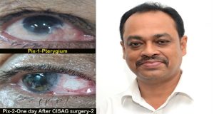
Eminent eye surgeon of Bokaro Dr. Sanjay Chowdhury has developed a new surgery technique to treat Pterygium— an eye problem. This new method of surgical technique or treatment is not just simple, easy and effective, but rather the probability of recurrence also decreases to a great extent.
The new study has been published in the prestigious peer-reviewed International Journal of Universal Surgery.
The study assumes importance considering that several treatment options have been tried out but recurrence of Pterygium is a big problem, he said.
The study was conducted over the number of patients and all of them showed considerable improvement. No complications or recurrence was observed; this process can also be combined successfully with modern day cataract surgery, he said.
Dr Sanjay, presently posted at Bokaro General Hospital (BGH) as an eye surgeon, run and managed by SAIL’s (Steel Authority of India Limited) Bokaro Steel Plant at Bokaro Steel City said, we have performed this technique on more than 65 patients during our study period of one year.
“Now this technique has become our routine procedure, more or less we are treating 2 to 3 Pterygium patients every day at BGH,” he added.
Explaining about the disease (Pterygium) Dr. Sanjay said it is an eye disease found mostly in the tropical countries which can have an adverse effect on vision. It can lead to severe scarring on the cornea, but this is rare.
“Scarring on the cornea needs to be treated because it can cause vision loss,” he added.
For minor cases, treatment usually involves eye drops or ointment to treat inflammation. In the more serious cases, treatment can involve surgical removal of the Pterygium, he said.
It is reported that it (Pterygium) increases with exposure to dusty windy weather since form the early days and can become cosmetic blemish; many treatment options have been tried but recurrence is a big problem, he said.
Bare sclera has the highest recurrence rate. To reduce this high incidence of recurrence covering the exposed sclera with grafts was introduced. It includes Conjunctival Auto Grafts (CAG) with or without stem cells, Amniotic Membrane Transplant (AMT).
Different adjuvant therapy ranging from beta irradiation to antimitotic agents was also started but all these were not devoid of complications due to cytotoxicity hence lost their popularity. Pterygium itself has origins from cytotoxic effect of UV radiation. Moreover unstable tear film and cytotoxic effects of UV radiation can incite vasculoendothelial growth factors (VEGF) production which can give rise to pterygium formation.
Histopathologically conjunctiva is not at fault in Pterygium. This is an elastotic degeneration of Subconjunctival tissues. So conjunctiva overlying Pterygium can be used for covering bare sclera which can give stable tear film and protect from UV radiation thus preventing recurrence.
Briefing his new technique Dr Sanjay said, while studying the pathology of the disease we have started a new surgical technique and named it as ‘Conjunctival in situ Autograft’ (CISAG).
The method includes surgical excision of conjunctiva from the body of Pterygium under local anesthesia, facilitated by injecting a small amount of sterile air with a very fine needle beneath the conjunctiva. This delineates the extent of healthy tissue and rest of the affected tissue dissected from the scleral bed and head of the pterygium is torn slowly from the cornea with a smooth forceps, said.
Limbal stem cells beneath the Pterygium also dissected with normal crescent knife and surface of the affected cornea is smoothened carefully with microdissection, he said.
The real advantages of this CISAG procedure are no healthy tissue is disturbed as in other autograft, no extra medicines are used like mitomycin which is an anticancer drug with various side effects, no extra tissue like the amniotic membrane or no suture or no glue is required, he said.
“Nasal side placement of limbal side of conjunctiva ensure growth if any at all in future will not affect cornea and thus vision will remain intact and no graft malposition was observed in this study”, added Dr Sanjay.
I profusely thank our DMS I/C Dr AK Singh for his guidance and inspiration to be innovative, economic and patient caring. I also thank my DNB students Dr. Sneha and Dr. Priti to be part of this team who have also mastered this technique. I am grateful to BGH eye operation theatre staffs and my sincere thanks to Dr Prakash Pandey I/C Pathology department of BGH for giving excellent histopathology backup for my work, he said.